Hemidesmosomes vs Desmosomes: Key Differences in Cell Adhesion
Have you ever wondered how your body's cells manage to stick together so perfectly? It's actually pretty fascinating when you think about it. At the heart of this cellular adhesion story are two critical structures: hemidesmosomes and desmosomes. These microscopic cellular components might sound like something out of a sci-fi novel, but they're incredibly important for maintaining the integrity of our tissues, especially our skin and organs.
Honestly, when I first learned about these structures in biology class, I found their names pretty intimidating. But once you break them down, they're not as complex as they sound. Think of them as specialized cellular "glue" that keeps our tissues from falling apart. Today, I'll walk you through the key differences between these two types of cell junctions, and trust me, by the end of this article, you'll have a much clearer picture of how our bodies maintain their structural integrity.
Before we dive into the nitty-gritty details, let me share a quick story. A few years ago, I was speaking with a dermatology student who explained how understanding these cellular structures helped her better comprehend certain skin conditions. She mentioned that patients with certain genetic disorders affecting desmosomes or hemidesmosomes could experience severe skin blistering. That's when it really hit me just how crucial these tiny structures are for our everyday health and wellbeing.
What Are Hemidesmosomes?
Let's start with hemidesmosomes – and yes, the prefix "hemi" means "half," which gives you a pretty good hint about their structure. These tiny cellular components are essentially specialized adhesion structures that connect the basal cells of epithelial tissue to the underlying basement membrane. Think of them as anchors that keep your skin cells firmly attached to the deeper layers of tissue beneath.
The structure of hemidesmosomes is quite remarkable. They're made up of several proteins working together in harmony. The key players here include integrins (specifically integrin α6β4), plectin, and BP180 (also known as BPAG2). Now, I know these names might sound like alphabet soup, but each component has a crucial role. Integrins act as transmembrane receptors that span the cell membrane, while plectin serves as a cytoskeletal linker protein, connecting the whole structure to intermediate filaments inside the cell.
What's particularly interesting about hemidesmosomes is that they come in two types: Type I and Type II. Type I hemidesmosomes are found in stratified epithelia like your skin, while Type II are simpler structures found in other tissues. From what I understand, these structures are incredibly important for maintaining skin integrity – without them, our skin would basically peel right off! It's amazing how such small structures can have such a massive impact on our body's function.
Understanding Desmosomes
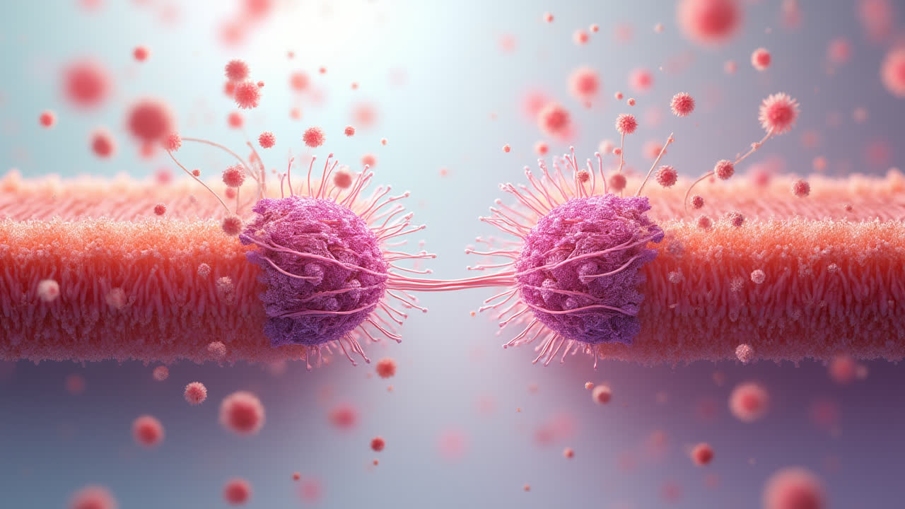
Now, let's talk about desmosomes, which are quite different from their "half" cousins. These structures are responsible for connecting adjacent cells to each other, rather than connecting cells to the basement membrane. You can think of desmosomes as molecular "spot welds" between cells – they create strong points of adhesion that help tissues withstand mechanical stress.
The molecular architecture of desmosomes is fascinating, though I'll admit it took me a while to wrap my head around it. These structures contain specialized proteins called cadherins – specifically desmogleins and desmocollins. These proteins extend from one cell to meet their counterparts from the neighboring cell, creating a strong intercellular bond. Inside the cell, these cadherin proteins connect to intermediate filaments through adapter proteins like plakoglobin and desmoplakin.
What I find particularly cool about desmosomes is where you find them. They're especially abundant in tissues that experience a lot of mechanical stress – like heart muscle, skin, and the lining of your digestive tract. Makes sense, right? These are areas that need extra reinforcement to handle all the stretching, pulling, and general wear and tear they experience daily. Without properly functioning desmosomes, these tissues would literally fall apart under stress!
Core Structural Differences
When comparing these two types of cell junctions, the structural differences become quite apparent. Hemidesmosomes, as their name suggests, look like half of a desmosome when viewed under an electron microscope. They're asymmetrical structures that connect cells to the extracellular matrix, while desmosomes are symmetrical structures that connect cells to other cells.
The protein composition is another major difference. Hemidesmosomes rely heavily on integrins as their primary adhesion molecules, while desmosomes use cadherins. This difference in adhesion proteins reflects their different functions – integrins are particularly good at binding to extracellular matrix proteins, while cadherins excel at cell-to-cell adhesion.
I remember being confused about this at first, but a helpful analogy from my professor cleared it up: Think of hemidesmosomes as anchors that tie a boat (the cell) to the dock (the basement membrane), while desmosomes are like ropes that tie multiple boats (cells) together. This simple visualization really helped me understand why these structures need different molecular components to perform their specific functions.
Functional Differences
The functional distinctions between hemidesmosomes and desmosomes are pretty significant. Hemidesmosomes serve as anchoring points that attach epithelial cells to the underlying connective tissue. This attachment is crucial for maintaining tissue integrity and preventing the separation of epithelial layers from their foundation. Without these structures, your skin would literally detach from the underlying tissues – not a pleasant thought!
Desmosomes, on the other hand, create strong cell-to-cell adhesions that allow tissues to function as cohesive units. They're particularly important in tissues that experience mechanical stress, providing the strength needed to prevent cells from being pulled apart. In cardiac muscle, for instance, desmosomes ensure that heart muscle cells stay connected during the powerful contractions of each heartbeat.
Here's something that really drives home the importance of these structures: certain genetic disorders affecting either hemidesmosomes or desmosomes can have devastating effects. Epidermolysis bullosa, which affects hemidesmosomes, causes severe skin blistering, while disorders affecting desmosomes can lead to conditions like arrhythmogenic right ventricular cardiomyopathy, a serious heart condition. It's sobering to think how these microscopic structures can have such profound impacts on human health.
Location and Distribution
The distribution of these cellular structures throughout the body is quite specific and relates directly to their functions. Hemidesmosomes are predominantly found in epithelial tissues, particularly in the basal layer of stratified epithelia like the skin and cornea. They're also present in other epithelial tissues including the respiratory tract and parts of the digestive system.
Desmosomes have a broader distribution and are found in tissues that need to withstand significant mechanical stress. They're abundant in the epidermis (the outer layer of skin), cardiac muscle, and the epithelial linings of various organs. One interesting tidbit I learned is that desmosomes are particularly numerous in tissues that experience frequent stretching or compression.
What's fascinating is how the distribution of these structures reflects the mechanical needs of different tissues. For example, the palms of your hands and soles of your feet have an especially high concentration of both hemidesmosomes and desmosomes. This makes perfect sense when you consider how much mechanical stress these areas endure daily. Nature really has thought of everything, hasn't it?
Clinical Significance
Understanding the clinical importance of hemidesmosomes and desmosomes opens up a whole new perspective on various diseases. Mutations affecting the proteins that make up these structures can lead to serious medical conditions. For instance, mutations in hemidesmosome components can cause different forms of epidermolysis bullosa, a group of inherited disorders characterized by fragile skin that blisters easily.
Autoimmune diseases can also target these structures. Pemphigus, for example, is an autoimmune condition where the body produces antibodies against desmosomal proteins, leading to severe skin blistering. Similarly, bullous pemphigoid involves autoantibodies targeting hemidesmosomal components. These conditions can be quite serious and require careful medical management.
From a therapeutic perspective, understanding these structures has led to better treatment approaches. Researchers are exploring ways to strengthen these cellular junctions or replace defective proteins in genetic disorders. Some experimental treatments even involve gene therapy to correct the underlying genetic defects. It's exciting to think about how our growing understanding of these basic cellular structures could lead to new treatments for previously untreatable conditions.
| Feature | Hemidesmosomes | Desmosomes |
|---|---|---|
| Primary Function | Cell-to-matrix adhesion | Cell-to-cell adhesion |
| Structure | Asymmetrical (half structure) | Symmetrical (complete structure) |
| Main Adhesion Proteins | Integrins (α6β4) | Cadherins (desmogleins, desmocollins) |
| Location | Basal layer of epithelial cells | Between adjacent epithelial cells |
| Key Components | Plectin, BP180, BP230 | Plakoglobin, desmoplakin |
| Associated Diseases | Epidermolysis bullosa, bullous pemphigoid | Pemphigus, arrhythmogenic cardiomyopathy |
| Connects To | Basement membrane/ECM | Adjacent cells |
| Found In | Skin, cornea, respiratory tract | Skin, heart muscle, GI tract |
Frequently Asked Questions
What happens when hemidesmosomes or desmosomes malfunction?
When these cellular structures malfunction due to genetic mutations or autoimmune attacks, serious medical conditions can develop. Hemidesmosome dysfunction can lead to skin blistering disorders like epidermolysis bullosa, where the skin becomes extremely fragile and separates from underlying tissues. Desmosome dysfunction can cause conditions ranging from severe skin disorders (pemphigus) to heart problems (arrhythmogenic cardiomyopathy), as cells lose their ability to properly adhere to each other.
Can damaged hemidesmosomes and desmosomes be repaired?
Yes, in many cases, damaged hemidesmosomes and desmosomes can be repaired through the body's natural healing processes. Cells continuously produce new proteins to maintain and repair these structures. However, in genetic disorders where the proteins themselves are defective, repair is more challenging. Current research is exploring gene therapy and protein replacement strategies to address these genetic conditions, though many treatments are still in experimental stages.
Why are these structures important for wound healing?
Hemidesmosomes and desmosomes play crucial roles in wound healing by maintaining tissue integrity during the repair process. During wound healing, cells need to migrate to close the wound while maintaining attachment to prevent tissue separation. These structures are dynamically regulated during healing - they must loosen to allow cell movement, then reform to restore tissue strength. Proper function of both types of junctions is essential for successful wound closure and the restoration of tissue barrier function.
Conclusion
Understanding the differences between hemidesmosomes and desmosomes reveals the incredible complexity of our cellular architecture. While both structures serve as cellular adhesion points, their distinct roles – with hemidesmosomes anchoring cells to the basement membrane and desmosomes connecting cells to each other – are fundamental to maintaining tissue integrity throughout our bodies.
These microscopic structures might seem insignificant at first glance, but as we've seen, they're absolutely critical for our health. From keeping our skin intact to ensuring our heart beats properly, hemidesmosomes and desmosomes work tirelessly behind the scenes to maintain the structural integrity of our tissues.
The ongoing research into these cellular junctions continues to provide insights that could lead to new treatments for various diseases. As our understanding grows, so does our appreciation for these remarkable molecular machines that keep us literally held together. Whether you're a student, healthcare professional, or simply curious about how your body works, understanding these fundamental cellular structures gives you a deeper appreciation for the intricate design of human biology.