SEM vs TEM: Essential Differences Between These Powerful Microscopy Techniques
Understanding Electron Microscopy: The Basics
When I first encountered electron microscopy during my research days, I was amazed at how these instruments could reveal structures invisible to conventional light microscopes. Both scanning electron microscopy and transmission electron microscopy use beams of electrons rather than light to create images of specimens, but they do so in fundamentally different ways.
Think of it this way: if a regular microscope is like looking at an object with a flashlight, SEM is like running your fingers over a surface to feel its texture, while TEM is more like shining a light through a thin slice of material to see what's inside. The difference between SEM and TEM isn't just technical—it determines what kind of information you can extract from your samples.
I remember spending weeks learning how to prepare samples properly for both techniques. Trust me, there's nothing quite like the frustration of realizing your sample prep went wrong after hours of work! But that's part of the journey when working with these sophisticated tools that can reveal the nanoscale world.
Before we dive deeper into their differences, let's get a clear understanding of what each of these microscopy techniques actually involves. After all, you can't appreciate the distinctions without first understanding what makes each one unique in its own right.
What is SEM (Scanning Electron Microscope)?
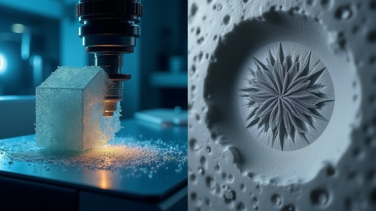
A Scanning Electron Microscope (SEM) works by scanning a focused beam of electrons across the surface of a specimen. When I first watched an SEM in action, it reminded me of an old television—both build images line by line, though an SEM does so with considerably more precision! As the electrons interact with atoms at the surface of the sample, they produce various signals that are detected to create a detailed image.
What makes SEM particularly useful is its ability to reveal the topography and composition of materials with remarkable clarity. The electrons emitted by the electron gun are accelerated toward the specimen by an electric field, and these interactions generate secondary electrons, backscattered electrons, and characteristic X-rays. Each of these signals provides different information about the specimen—secondary electrons primarily reveal morphology and topography, while backscattered electrons highlight compositional contrasts.
I've always been impressed by how SEM can provide a three-dimensional appearance of specimens, making it easier to understand surface structures. And the depth of field? Simply incredible. I once examined a butterfly wing under SEM, and the intricate scales and ridges appeared as if I were flying over a landscape of mountains and valleys.
One major advantage of SEM over other microscopy techniques is its versatility in sample types. It can analyze various materials including biological samples, polymers, metals, and ceramics. For advanced analytical capabilities, SEM can be equipped with accessories like Energy-dispersive X-ray spectroscopy (EDS) to identify elemental composition. This versatility makes SEM an indispensable tool across disciplines—from forensics to nanotechnology and semiconductor manufacturing.
However, SEM isn't without limitations. Samples generally need to be conductive, which often means coating non-conductive specimens with a thin layer of metal like gold or carbon. And while SEM provides excellent surface details, it can't peek inside your samples the way TEM can. It's a bit like being able to examine the cover of a book in exquisite detail without being able to read the pages inside.
What is TEM (Transmission Electron Microscope)?
Transmission Electron Microscopy (TEM) operates on a different principle altogether. Instead of scanning the surface, TEM sends electrons through an ultra-thin sample. The first time I saw a TEM image, I was stunned by its ability to reveal internal structures at a near-atomic level. It's like having X-ray vision, but with substantially better resolution!
As electrons pass through the specimen in TEM, they interact with the sample's atoms, being scattered or absorbed depending on the material's composition and thickness. These interactions produce various signals, including transmitted electrons that form the primary image and diffracted electrons used for structural analysis through techniques like electron diffraction. This ability to "see through" samples makes TEM particularly valuable for studying internal structures and features.
The resolution capabilities of TEM are truly mind-boggling. Modern TEMs can achieve resolutions better than 0.1 nanometers, allowing researchers to visualize individual atoms within a sample. I remember the first time I saw an image of a crystal lattice where you could actually count the atoms—it was a profound moment that connected theory with observable reality.
Like SEM, TEM can be combined with analytical techniques such as energy-dispersive X-ray spectroscopy (EDS) to determine elemental composition. But TEM goes further by allowing analysis of crystallographic information through electron diffraction patterns—a capability that's proven invaluable in materials science and semiconductor research.
The catch with TEM? Sample preparation is notoriously difficult and time-consuming. Specimens must be extremely thin—typically less than 100 nanometers—for electrons to pass through. Achieving this thinness requires specialized techniques like ultramicrotomy, ion milling, or focused ion beam (FIB) preparation. I've spent entire days preparing a single TEM sample, only to have it tear or contaminate during the final stages. It's painstaking work, but the resulting images make it worthwhile.
Similarities Between SEM and TEM
Despite their operational differences, SEM and TEM share important commonalities. Both techniques rely on electron beams rather than light, allowing them to overcome the resolution limitations of optical microscopes. This shared foundation means they both require vacuum systems to prevent electrons from being scattered by air molecules. I've experienced the frustration of vacuum pump failures on both types of instruments—nothing quite like watching your research grind to a halt while waiting for repairs!
Both microscopy methods offer high-resolution images far beyond what's possible with optical microscopes. They also require specialized sample preparation, though the specific requirements differ substantially. Additionally, both SEM and TEM typically require conductive or specially treated samples, and both can be equipped with analytical capabilities like EDS for elemental analysis.
In my experience, mastering either technique requires significant training and practice. The learning curve is steep, but once you've developed proficiency, you gain access to an incredible realm of imaging possibilities. Both techniques have transformed numerous scientific fields by revealing structures and processes previously hidden from human observation.
Key Differences Between SEM and TEM
Now, let's explore the crucial differences that determine which technique is appropriate for a particular research question. These distinctions go beyond simple operational variations—they fundamentally affect what kinds of samples you can examine and what information you can extract.
The most fundamental difference between these techniques lies in their imaging approach: SEM scans the surface of a specimen with electrons to create an image of its topography, while TEM transmits electrons through an ultra-thin specimen to reveal its internal structure. This core distinction drives all other differences between the methods.
| Feature | SEM (Scanning Electron Microscope) | TEM (Transmission Electron Microscope) |
|---|---|---|
| Basic Principle | Scans surface with electron beam | Transmits electrons through specimen |
| Image Type | Surface morphology and topography | Internal structure and ultrastructure |
| Sample Preparation | Relatively simple; coating with conductive material | Complex; requires ultrathin sectioning |
| Resolution | 1-20 nm (lower resolution) | 0.1-0.5 nm (higher resolution) |
| Depth of Field | Large (3D-like appearance) | Small (flat appearance) |
| Sample Thickness | Can be thick; bulk specimens | Must be extremely thin (<100 nm) |
| Magnification Range | 10x to 100,000x | 50x to 5,000,000x |
| Cost | Generally lower | Generally higher |
Beyond these technical specifications, the choice between SEM and TEM often comes down to the research question at hand. If you're interested in surface details, textures, or compositional analysis, SEM is typically the go-to method. If you need to visualize internal structures, crystallographic information, or achieve atomic-level resolution, TEM is the better choice.
In my laboratory work, I've found that these techniques often complement each other. For instance, when investigating nanoparticles, we might use SEM to characterize their size distribution and surface morphology, then turn to TEM to examine their internal crystal structure and composition. This complementary approach provides a more comprehensive understanding than either technique alone could offer.
Applications of SEM and TEM in Various Fields
The applications of electron microscopy span virtually every scientific discipline. In materials science, SEM helps characterize surface properties of alloys, ceramics, and polymers, while TEM reveals crystal structures and defects. The semiconductor industry relies heavily on both techniques for quality control and failure analysis—I recall assisting a company in identifying nanoscale defects in microchip production using TEM analysis.
In biological sciences, SEM provides detailed images of cells, tissues, and organisms—revealing the intricate surface structures of everything from pollen grains to insect eyes. TEM, meanwhile, has been fundamental in understanding cellular ultrastructure, visualizing viruses, and studying protein structures. Medical research benefits from both techniques for pathology, tissue engineering, and pharmaceutical development.
Forensic science has also embraced electron microscopy, with SEM being particularly useful for analyzing gunshot residue, paint chips, and fiber evidence. Environmental sciences use both techniques to study particulate pollution, soil microstructures, and mineral compositions. Even the art world employs electron microscopy for authentication and conservation of paintings and artifacts.
One particularly fascinating application I've witnessed is in archaeology, where SEM has been used to examine the microstructure of ancient ceramics to determine firing temperatures and manufacturing techniques. Similarly, TEM has helped analyze the composition of ancient glass and metals, providing insights into historical production methods.
Frequently Asked Questions About SEM and TEM
Why is SEM often preferred over TEM in many applications?
SEM is frequently preferred over TEM for several practical reasons. First, SEMs are generally more affordable, making them accessible to a wider range of laboratories and institutions. Sample preparation for SEM is considerably simpler and less time-consuming than the ultrathic sectioning required for TEM. Additionally, SEM can accommodate larger samples and provides excellent surface detail with a good depth of field, creating images with a three-dimensional appearance. For many applications where internal structure isn't the primary concern, these advantages make SEM the more efficient and cost-effective choice.
Can electron microscopes produce color images?
Neither SEM nor TEM naturally produces color images because electrons don't have color properties like photons (light) do. The grayscale images from electron microscopes represent differences in electron density or scattering rather than actual colors. However, images are often artificially colorized after acquisition for several reasons: to highlight specific features, to represent elemental or compositional differences (especially when combined with techniques like EDS), or simply to make structures more visually distinguishable. These colorized images are useful for presentation and interpretation but do not represent actual colors in the specimen.
What recent advances have improved electron microscopy capabilities?
Electron microscopy has seen remarkable advances in recent years. For TEM, developments like aberration correction have dramatically improved resolution, allowing for true atomic-scale imaging. Cryo-electron microscopy (cryo-EM) has revolutionized structural biology by enabling visualization of proteins in their native states, leading to a Nobel Prize in 2017. For SEM, environmental chambers now allow imaging of hydrated specimens, while focused ion beam (FIB) integration enables precise sample preparation and 3D reconstruction. Additionally, advances in detector technology have improved signal-to-noise ratios, and automated data collection and analysis software have made these powerful tools more accessible to non-specialists, expanding their applications across scientific disciplines.
Conclusion: Choosing Between SEM and TEM
The choice between SEM and TEM ultimately depends on your specific research question, sample characteristics, and available resources. SEM excels at revealing surface details with excellent depth of field and relatively straightforward sample preparation. It's ideal for examining surface morphology, topography, and composition. TEM, with its superior resolution and ability to visualize internal structures, is unmatched for applications requiring atomic-level detail or analysis of internal features.
In many research settings, these techniques complement rather than compete with each other. The combined insights from both methods often provide a more comprehensive understanding than either alone. As technology continues to advance, we're seeing increasingly sophisticated hybrid systems that combine features of both techniques, along with other analytical methods.
Whether you're studying nanoparticles, biological specimens, advanced materials, or ancient artifacts, understanding the fundamental differences between these powerful microscopy techniques is essential for selecting the right tool for your scientific investigation. The remarkable capabilities of both SEM and TEM continue to push the boundaries of what we can observe and understand about our world at the nanoscale.